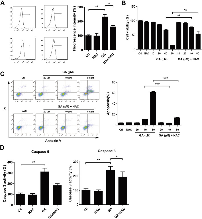
Fig. 4.
NAC treatment attenuated GA-mediated HaCaT keratinocytes growth inhibition and apoptosis. (A,D) HaCaT keratinocytes were seeded into 6-well plates and treated with GA (80 μM) and/or NAC (5 mM) for 24 h. Cells were harvested and incubated with DCFH-DA for detection of ROS levels by flow cytometry. Caspase 9 and 3 activities were determined using caspase activity assay kits. *P < 0.05. **P < 0.01. (B, C) HaCaT keratinocytes were treated with GA (indicated concentration) and/or NAC (5 mM) for 24 h. Cell viability was measured using CCK-8 assay. Cell apoptosis was analyzed by flow cytometry. **P < 0.01. ***P < 0.001

