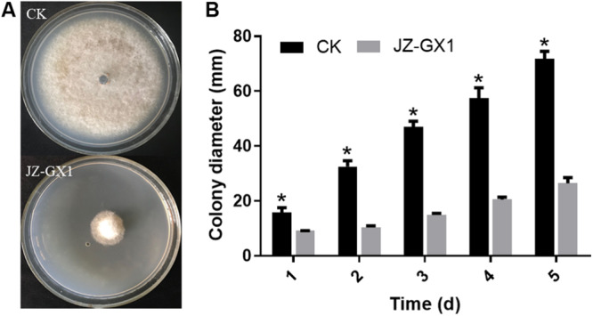
FIGURE 1.

Effect of R. aquatilis JZ-GX1 on the mycelial growth of C. gloeosporioides. (A) Colony morphology of C. gloeosporioides in vitro. (B) Colony diameter of C. gloeosporioides. Vertical bars represent the standard deviation of the average. One-way ANOVA analysis was performed and Duncan’s post hoc test was applied. Asterisks indicate statistically significant differences (p < 0.05) among treatments.
