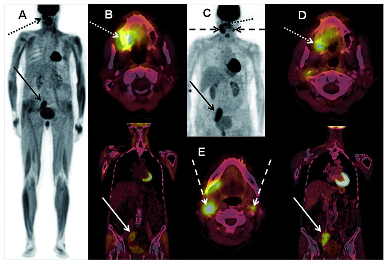
Figure 3.
Images of a 63-year old female patient with histologically confirmed extramedullary (EM) manifestation of acute myeloid leukemia (AML) in the oral cavity (dotted arrows) who underwent intensive induction chemotherapy. (A) Maximum intensity projection (MIP) and (B) fused multiplanar reconstruction (MPR) of the pre-therapeutic 18Fluorodesoxy-glucose positron emission tomography/computed tomography (18FDG-PET/CT). Maximum standardized uptake value (SUVmax) was 9.1. 18FDG-PET detected a further right iliac EM AML (SUVmax 5.6; continuous arrows). (C) MIP of the post-therapeutic follow up 18FDG-PET/CT confirming the slightly regressive EM AML of the oral cavity (SUVmax 7.4) but also the progressive right iliacal EM AML (SUVmax 8.1). (D) MPR of this scan. New bicervical EM AML (dashed arrows) was also detected (E), see also (C) (SUVmax up to 9.5).

