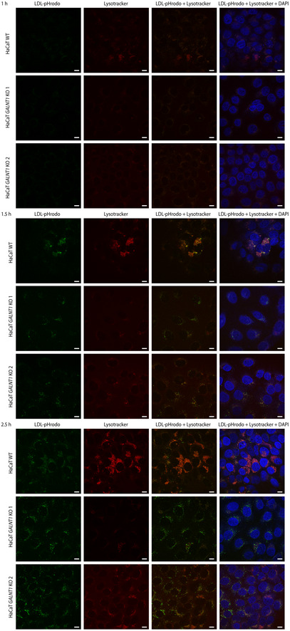
Figure EV3. LDL trafficking in HaCaT WT and GALNT1 KO cells.

Cells grown on coverslips were cultured in media with lipoprotein‐depleted serum for 24 h, followed by pulsing with Lysotracker Red DND‐99 (−1 h) and LDL‐pHrodo (0 h). Coverslips were fixed at 1 h (upper panels), 1.5 h (middle panels), and 2.5 h (lower panels) and imaged using confocal microscopy. Scale bar—10 μm.
