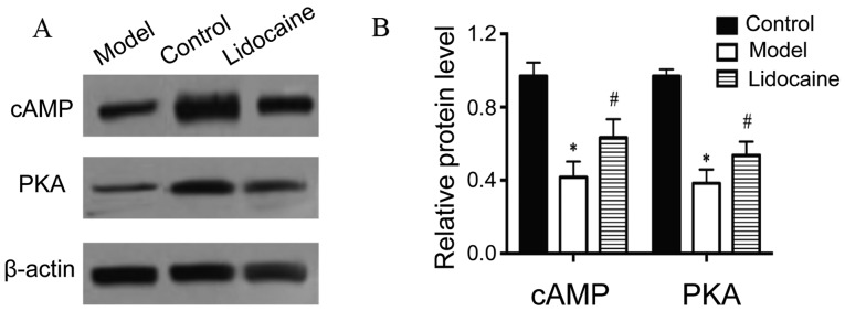
Figure 4.
Protein levels of cAMP and PKA in the cerebral tissues of rats by western blotting. (A) Western blotting bands. (B) Statistical graphs of the bands. *P<0.05, model group vs. control group; and #P<0.05, lidocaine group vs. model group. cAMP, cyclic adenosine monophosphate; PKA, protein kinase A.

