
Fig. 8. Activation of AKT and ERK signaling by FGF9 and FGF10 in lung explants lacking mesenchymal FGFR1 and FGFR2.
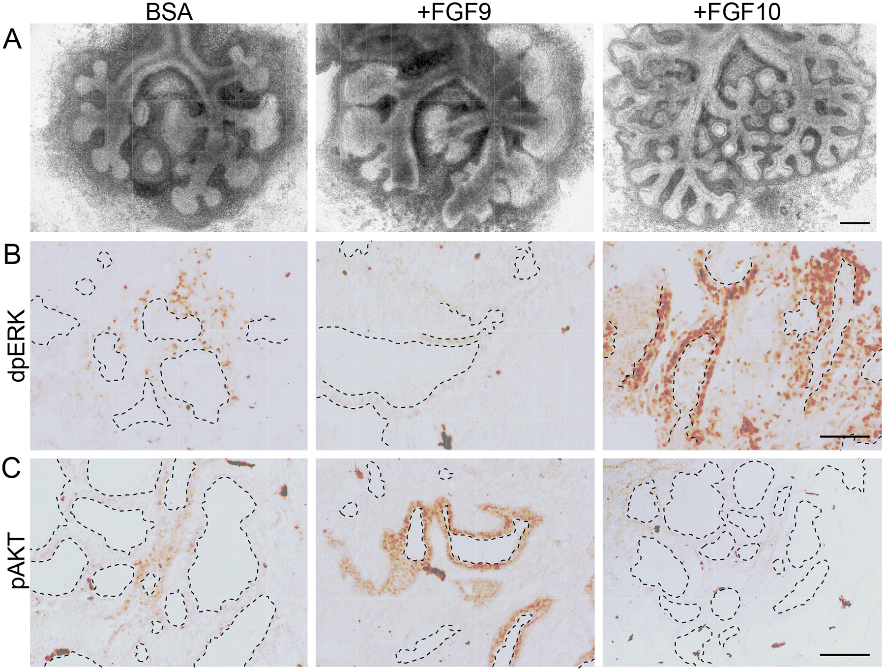
(A) Images of E11.5 whole lung explants from Dermo1-FGFR1/2-KO mice cultured for 48 h and treated with BSA, FGF9, or FGF10. (B, C) Sections of lung explants treated as in (A) immunostained for doubly phosphorylated (active) ERK (dpERK, B) and phosphorylated (active) AKT (pAKT, C). All images are representative of at least 3 embryos. Dashed lines outline ductal lumens. Scale bars, 200 μm (A); 100 μm (B and C).