Figure 2.
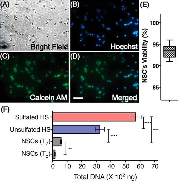
Cytocompatibility and proliferation of NSCs encapsulated in HS hydrogels. NSCs were stained for nuclei (Hoechst) and live cell (Calcein AM) detection after culturing for 24 h. A bright field image and an overlay of these images are also presented (A–D). Scale, 50 μm. (E) Viability of NSCs. (F) Total isolated DNA from NSCs grown on Geltrex coated culture plates (T0, day 0 and T7, day 7) and from NSCs encapsulated in the sulfated (14)-HS and unsulfated (13, control)-HS hydrogel. Statistical differences observed between groups are represented by **** indicating P < 0.001 and ** indicating P < 0.01.

