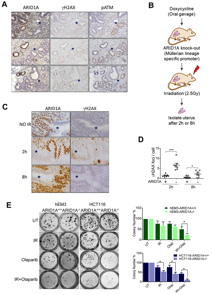
Fig. 1. ARID1A deficiency results in sustained DSBs and sensitizes cells to irradiation.
(A) Immunohistochemical staining patterns of ARID1A, γH2AX, and phosphorylated-ATM (s1981) in complex atypical hyperplasia (CAH) of endometrium. ARID1A-loss of expression areas are marked with blue stars. Representative images are shown. (B) Schematic illustration of the experimental procedure. (C) Immunohistochemical staining of ARID1A and γH2AX in endometrial tissue of ARID1Aflox/flox mice. Mice were treated with doxycycline for one week to induce deletion of ARID1A, and were sacrificed for analysis at the indicated time points after irradiation (2.5 Gy). ARID1A-loss areas are marked with blue stars. Representative images are shown. (D) Quantitation of γH2AX foci per endometrial epithelial cell. Data are presented as mean ± SEM; more than 500 cells were analyzed per group. Mann-Whitney test (two-tailed) was used to calculate significance; *p<0.05, ***p<0.001. (E) Clonogenic formation visualized by crystal violet staining on day 7 post-irradiation. Colony numbers were quantified and plotted (right panels). Data are presented as mean ± SEM, n=6; *p<0.05.

