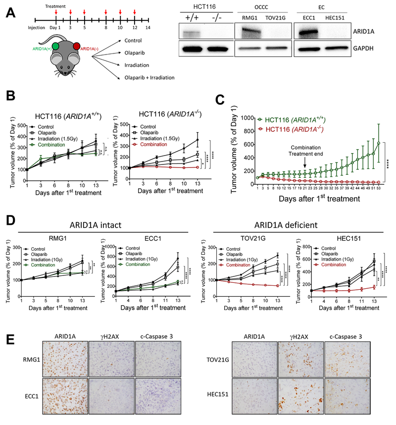
Fig. 4. Combination of PARP inhibitor and ionizing radiation inhibits growth and induces apoptosis in ARID1A-deficient tumor xenografts.
(A) (left) Schematic of treatment regimen in mice. Immunocompromised athymic nu/nu mice were inoculated with indicated human tumor cells, and 10–14 days later, mice were randomly stratified into 4 treatment groups. Olaparib (50 mg/kg) and/or irradiation (1–2 Gy) were administrated 3 times per week. (Right) Immunoblots performed to assess ARID1A protein expression in each cell line. (B) Tumor volume of HCT116-ARID1A+/+ and ARID1A−/− xenografts monitored for 2 weeks. Data are presented as mean ± SEM, n=5. Mann-Whitney test (two-tailed) was used to calculate the statistical significance between two comparison groups on day 13; *p<0.05, ****p<0.0001. (C) Relative tumor volume measured in mice inoculated with HCT116 cancer cells and treated with olaparib and ionizing radiation (2 Gy) combined therapy for three weeks. Tumor growth was monitored until day 53. Data are presented as mean ± SEM, n=4; ****p<0.0001. (D) Tumor volume measured in different groups. RMG1 and ECC1 are ARID1A wild-type and express ARID1A protein while TOV21G and HEC151 harbor inactivating mutations and lose ARID1A protein expression. Data are presented as mean ± SEM, n=5; *p<0.05, **p<0.01, ***p<0.001, ****p<0.0001. (E) Immunohistochemical staining of ARID1A, γH2AX, and cleaved caspase-3 in tumor xenografts excised from mice treated with olaparib and ionizing radiation (2 Gy).

