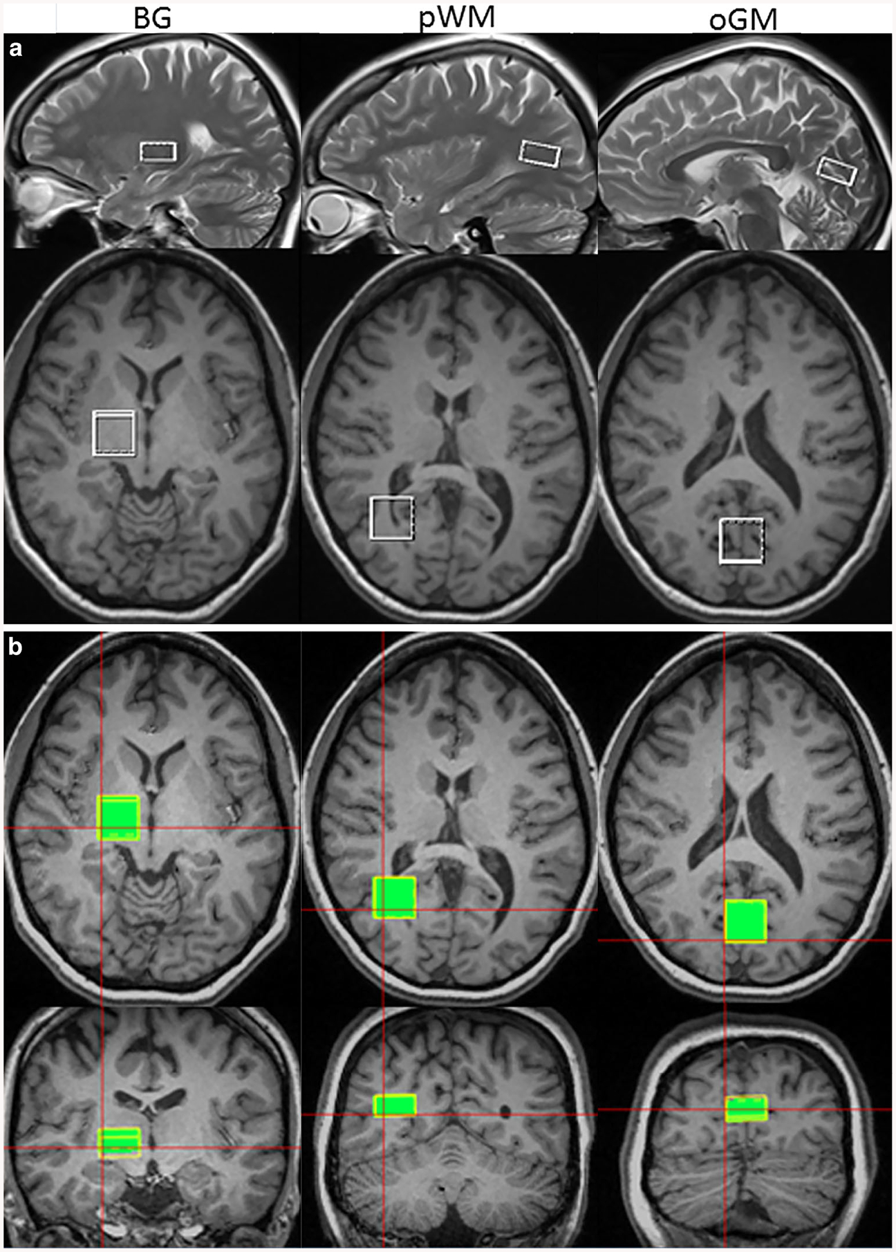
Fig. 2.

Locations of the ROIs in basal ganglia (BG), parietal white matter (pWM) and occipital grey matter (oGM) drawn on T1-weighted images in axial (2nd and 3rd rows), sagittal (upper row), and coronar (lower row) sections, shown for a the SVS acquisition and b the integrated wbMRSI measurement
