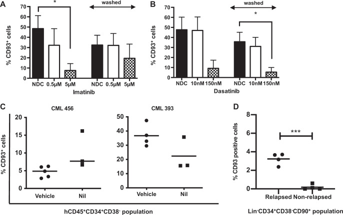
Fig. 5. TKIs downregulate CD93, but do not eliminate CD93+ LSC even after prolonged in vivo therapy.
CP-CML cells (n = 3) were thawed and sorted into an LSC population before culturing in SFM plus physiological growth factors with and without Imatinib (a) or Dasatinib (b). Following culture for 24 h, half of the samples were analyzed by flow cytometry to assess CD93 expression. The rest were thoroughly washed to completely remove TKI and re-cultured in SFM supplemented with physiological growth factors for a further 24 h prior to flow cytometry analysis. c A nilotinib-treated PDX model was used to determine CD93+ cells in the hCD45+CD34+CD38− population by flow cytometry. This demonstrates persistence of CD93 expression despite TKI treatment in two patient samples. d CD93 expression as determined by flow cytometry in eight patients with prolonged TKI treatment. Within patients who relapsed post TKI discontinuation, a small lin−CD34+CD38−CD90+CD93+ population could be identified, compared with patients who did not demonstrate a molecular relapse.

