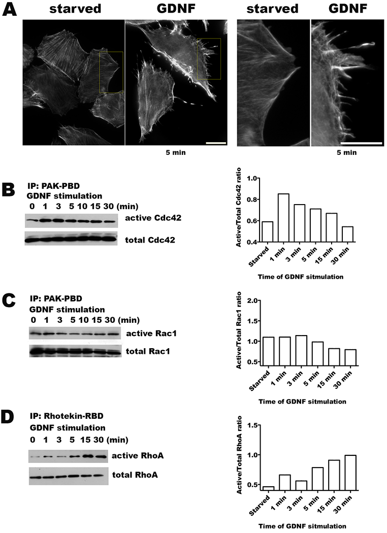
Figure 1. Activation of Rho GTPases by glial cell line-derived neurotrophic factor (GDNF).

A. Serum starved MiaPaCa2 cells were exposed to 100 ng/ml of GDNF for 5 minutes and then fixed in formaldehyde. Actin filaments were visualized with rhodamine-phalloidin staining, demonstrating the development of microspikes, filipodia, and stress fibers. Serum starved cells not exposed to GDNF were used as a control. (Scale bar = 15 μm) B-D. Western blots of GST pull-down assays assess for changes in the active, GTP-bound forms of Cdc42, Rac1, and RhoA in response to exposure to GDNF (100 ng/ml) with corresponding quantification by densitometry.
