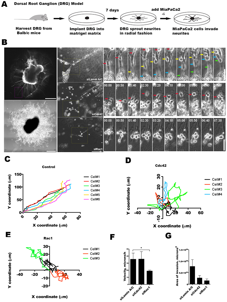
Figure 5. Cdc42 regulates the direction of cancer cell migration along nerves.

A. Dorsal root ganglia (DRG) were harvested from mice, grown in Matrigel, and co-cultured with MiaPaCa2 cancer cells as an in vitro model to explore cancer and nerve interactions. B. DRG implanted in Matrigel develops axonal projections or neurites, as imaged by immunofluorescence confocal microscopy using Tuj1 antibody (scale bar = 200 μm, left upper panel), or phase contrast microscopy, (scale bar = 400 μm, left lower panel). Representative MiaPaCa2 cells treated with siLaminA/C (upper central panel), siCdc42 (mid central panel), or siRac1 (lower central panel) are examined when associated with DRG neurites (scale bar = 200 μm). High power, sequential, time-lapse micrographs are shown (scale bar = 100 μm, right panels). Control siLaminA/C (right upper panels), siCdc42 (right mid panels), and siRac1 (right lower panels) MiaPaCa2 cells are depicted with colored arrows marking the migrating cells. C-E. Coordinate graphs depicting the single cell migration trajectories of control siLaminA/C (C), siCdc42 (D), and siRac1 (E) MiaPaCa2 cells. The siLaminA/C cells demonstrate consistent migration along the direction of the DRG neurites. The siCdc42 and siRac1 cells both exhibit a loss of directional migration. F. The average speed of single cell migration was calculated for the siLaminA/C, siCdc42, and siRac1 groups. Speed was maintained in the siLaminA/C and siCdc42 cells, but was significantly diminished in the siRac1 cells. G. The average area of DRG invasion was measured for siLaminA/C, siCdc42 and siRac1 cells after 48 hours of co-culture. Silencing of Cdc42 (p=0.08) or Rac1 (p=0.07) as compared with siLamin A/C resulted in a trend towards a decrease in the area of PNI.
