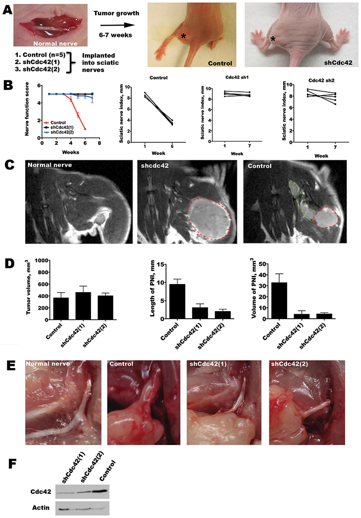
Figure 6. Cdc42 regulates perineural invasion in vivo.

A. An in vivo model of PNI involves the implantation of cancer cells into murine sciatic nerves. Stable MiaPaCa2 cell lines were generated with shRNA targeting Cdc42 or empty vector transfected controls. Representative images of mice 6 weeks after tumor implantation show left hind limb paralysis in the control tumor group, but normal limb function in the shCdc42 groups. Asterisks indicate the location of the sciatic nerve tumor. B. Sciatic nerve function was assessed by both the sciatic nerve function score (hind limb function), and the sciatic nerve index (hind paw width). Mean scores for sciatic nerve function score show a significant decline over 6 weeks for the control group (p<0.05, t-test), but not for the shCdc42 groups (p=NS, t-test). Similarly, the control group showed a significant decline in sciatic nerve index (p<0.05, t-test), but not for the shCdc42 groups (p=NS, t-test). To account for differential growth rates, comparisons were made between equivalent tumor volumes between week 6 data for the control group, and week 7 in the shCdc42 groups. C. Magnetic resonance images (MRI, T1 with gadolinium) are shown of a representative, non-injected, sciatic nerve (no tumor), an shCdc42 sciatic nerve tumor (lacking PNI), and a control sciatic nerve tumor (with PNI). The primary tumor is outlined in red, and the perineural component is outlined in green. D. Quantification of the primary tumor volume, the length of PNI, and the volume of tumor in the region of PNI by MRI. Comparisons were performed between week 6 for the control group, and week 7 in the shCdc42 groups when mean volumes were similar (p=NS, t-test). This comparison at different time points accounts for differences in tumor growth rate. Both the length of PNI and volume of tumor in the PNI were significantly reduced in both shCdc42 groups as compared with control (p<0.05 for both comparisons, for both shCdc42(1) and shCdc42(2), t-test). E. In-situ image of a surgically exposed sciatic nerve in a non-injected mouse, with control at week 6, and shCdc42(1) and shCdc42(2) tumors at week 7. Only the control has significant sciatic nerve thickening, consistent with perineural invasion, although all tumor groups developed equivalent primary tumor volume. F. Western blot of shCdc42(1) and shCdc42(2) MiaPaCa2 cell lines shows Cdc42 depletion as compared with control.
