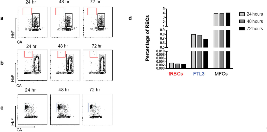
Fig. 2: Fetal RBC and maternal F cell stability at 24, 48 and 72 hours after staining.
(a) Adult nulliparous female RBCs showing the maternal F cell fraction (MFC, black gate) and no fetal RBCs (fRBCs, red gate). (b) Blood from the same adult subject plus cord blood (diluted at 1:10,000). (c) Fetaltrol (FTL3, blue gate) sample analyzed at 24, 48 and 72 hours after staining showed stability of fetal RBCs. (d) Quantitation of fetal RBC and maternal F cell from panel b and fetaltrol from panel c.

