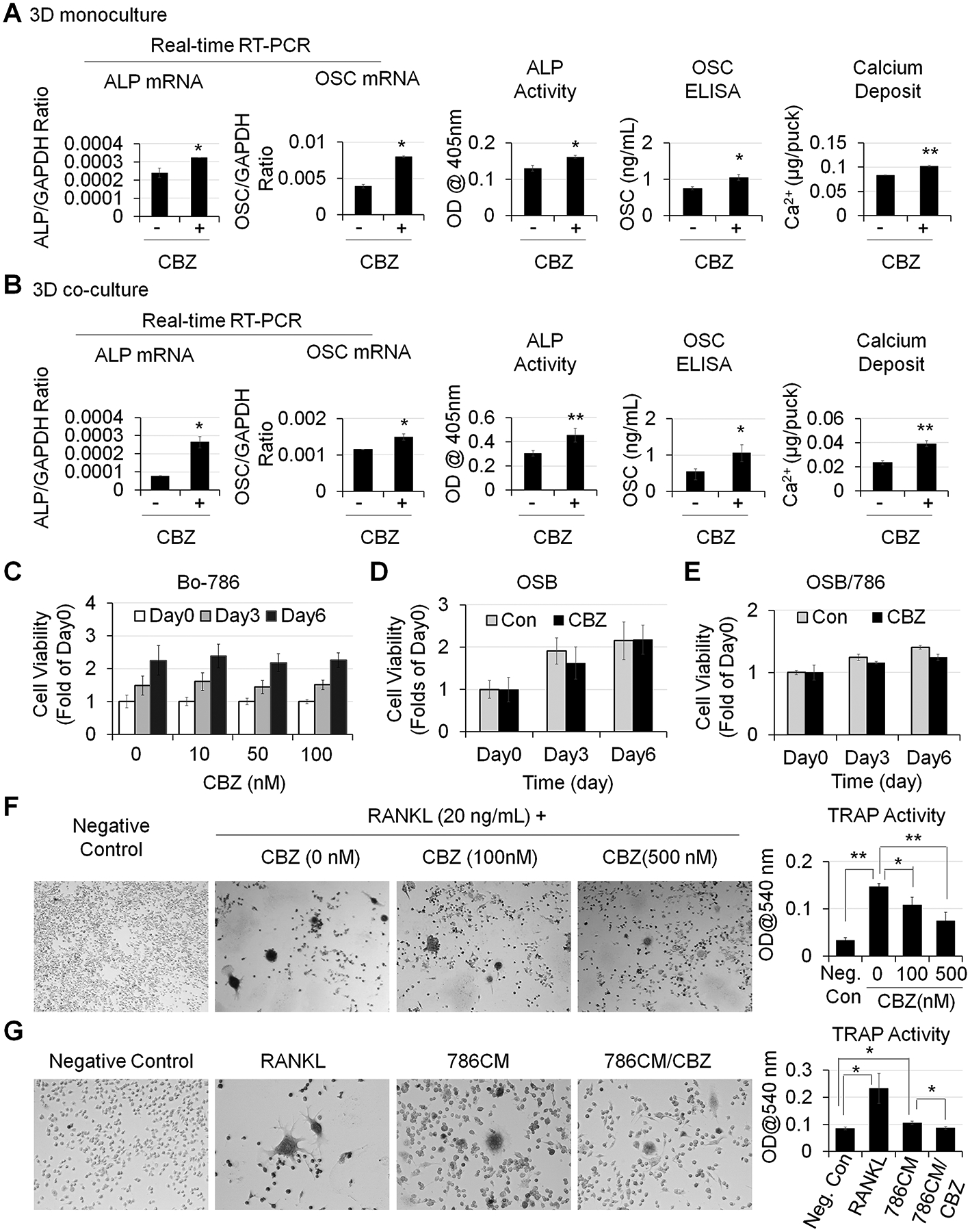
Figure 4. CBZ effects on OSB and osteoclasts in vitro.

A) OSB in 3D monoculture and B) OSB in 3D co-cultured with Bo-786 cells were treated with or without CBZ (100 nM) in ODM for 6 days. ALP and OSC mRNA and protein levels and calcium deposits were determined as in Fig. 3. C) Bo-786 cells cultured in 3D gel were treated with or without CBZ at various concentrations from 0 to 100 nM. Cell viability was determined by PrestoBlue assay. D) OSB and E) OSB co-cultured with Bo-786 cells in 3D gel were treated with or without CBZ (100 nM). Cell viability was determined by PrestoBlue assay. F) RAW264.7 cells were treated with RANKL at 20 ng/mL with or without CBZ (100 nM, 500 nM) for 5 days. Differentiation of RAW264.7 cells was determined by TRAP staining and TRAP activity assay. G) RAW264.7 cells were treated with 10-fold concentrated Bo-786 CM with or without CBZ (100 nM) for 5 days. RAW264.7 cells treated with RANKL (50 ng/mL) were used as positive control. Differentiation of RAW264.7 cells was determined by TRAP staining and TRAP activity assay. *: P < 0.05; **: P < 0.01.
