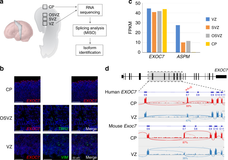
Fig. 3. EXOC7 is highly expressed in developing cortex.
(a) Diagram of cortical section of human fetal cortex indicating locations of RNAscope imaging in (b) and RNA sequencing in (c). (b) RNAscope imaging of fetal human cortex shows EXOC7 expression in VZ and OSVZ, two progenitor zones, and in CP, the location of postmitotic neurons. (c) EXOC7 is highly expressed in developing human cortex and shown in comparison with ASPM. Expression levels measured based on RNA sequencing.33 (d) RNA sequencing data from developing human fetal cortex (GW15) and mouse cortex (E14.5) showing differential inclusion of exon 7 in CP vs. VZ. FPKM fragments per kilobase of transcript per million mapped reads.

