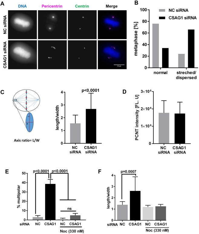
Fig. 4.
Mitotic spindle microtubules are essential for CSAG1 depletion-induced changes in the distribution of pericentrin at metaphase. (A) Immunofluorescence images of HeLa cells labeled for centrin and pericentrin that were transfected with control siRNA (top set of panels) or CSAG1 siRNA (bottom set of panels). Cell nuclei were stained with DAPI (DNA), merged images are shown in the last column. (B) The fraction of bipolar metaphase cells with abnormal or dispersed, or normal (i.e. oval) distribution of pericentrin determined in cells described in A. A total of >100 cells was analyzed for each sample. (C) Schematic, showing the method to quantify the axis ratio (length/width) and the graph shows the ratios calculated. More than 50 cells were analyzed for each group. A Mann–Whitney test was used for statistical analysis. (D) Total amount of pericentrin was measured for the sum of z-stack images derived from cells as described in B. (E,F) Multipolarity (E), and pericentrin shape and/or distribution (F) was evaluated in CSAG1-depleted cells that had been treated with or without Nocodazole. CSAG1 depletion causes abnormal pericentrin shape and/or distribution in bipolar metaphase cells but does not alter the amount of pericentrin at poles. The redistribution requires intact microtubules. Mann–Whitney test was used for statistical analysis. Error bars represent +s.d.; ns, not significant.

