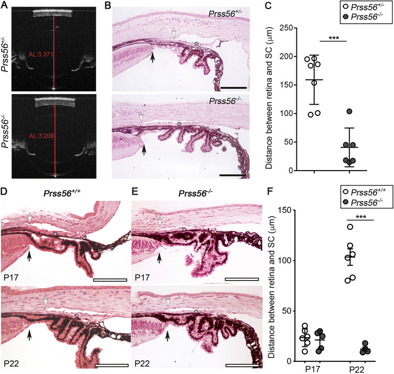
Fig. 1.
Prss56 knockout mice show altered positioning of ocular angle structures. (A) Representative OCT images showing reduced ocular size in Prss56−/− compared to Prss56+/− mice as previously described (Paylakhi et al., 2018). Shown are eyes from 2-month-old mice. The red lines indicate ocular axial length (AL). (B) Representative H&E stained ocular sections from Prss56−/− and control Prss56+/− mice showing altered organization of ocular angle structures in Prss56−/− mice that is characterized by a posterior shift in the position of the TM (asterisks) and adjoining Schlemm's canal (white arrows) relative to the iris and ciliary body, resulting in angle-closure. Black arrows indicate peripheral retina. (C) The distance between the peripheral retina and the posterior edge of the Schlemm's canal is significantly reduced in Prss56−/− eyes compared to control Prss56+/− eyes. (D,E) Representative H&E stained ocular sections from Prss56+/+ (D) and Prss56−/− (E) mice at different postnatal developmental stages showing that the distance between the peripheral edge of the retina (black arrows) and the posterior edge of the Schlemm's canal (white arrows) is narrow at P17 and is markedly increased at later time points (P22) in Prss56+/+, but not in Prss56−/− mice, leading to an anterior positional shift of ocular drainage tissues and open angle configuration. Asterisks indicate position of the TM. In C, n=7 Prss56+/− eyes, n=6 Prss56−/− eyes. In F, n=6 P17 Prss56+/+ eyes, n=5 P17 Prss56−/− eyes, n=6 P22 Prss56+/+ eyes, n=4 P22 Prss56−/− eyes. Data are mean±s.d., ***P<0.001, Student's t-test. Scale bars: 100 µm.

