Fig. 3.
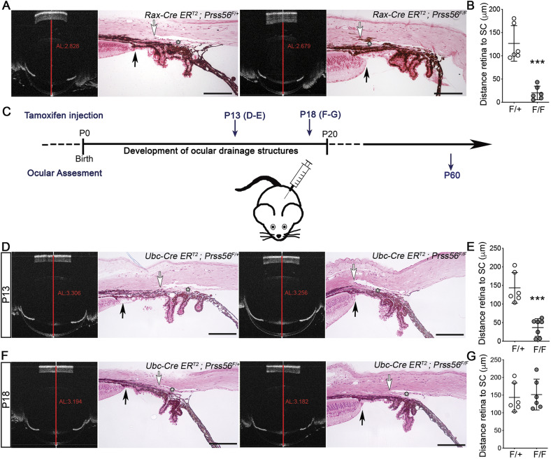
Spatiotemporal requirement of PRSS56 for proper positioning of ocular angle structures. (A,B) Representative OCT images and H&E-stained ocular sections (A) from control Rax-CreERT2;Prss56F/+ and Rax-CreERT2;Prss56F/F mice following tamoxifen injection at P8, a time point that coincides with the final differentiation of Müller cells. As described previously (Paylakhi et al., 2018), Rax-CreERT2;Prss56F/F mice (F/F) show reduced ocular axial length (AL, red lines on OCT images) compared to control Rax-CreERT2;Prss56F/+ mice (F/+). In addition, Rax-CreERT2;Prss56F/F mice exhibit a posterior shift in the position of the TM (asterisk) and adjoining Schlemm's canal relative to the iris and ciliary body, leading to a shorter distance between the peripheral retina (black arrows) and the posterior edge of the Schlemm's canal (white arrows) (quantified in B) and angle closure. (C) Schematic illustrating the tamoxifen injection paradigm for conditional inactivation of Prss56 at P13 and P18. (D,E) Representative OCT images and H&E-stained ocular sections (D) showing reduced ocular axial length as previously described (Paylakhi et al., 2018), and abnormal organization of ocular angle structures characterized by a posterior shift in the position of the TM (asterisk) and adjoining Schlemm's canal relative to the iris and ciliary body, leading to a shorter distance between the peripheral retina (black arrows) and the posterior edge of the Schlemm's canal (white arrows) (quantified in E) and angle closure in Ubc-CreERT2;Prss56F/F mice (F/F) following tamoxifen injection at P13 compared to control Ubc-CreERT2;Prss56F/+ mice (F/+). (F,G) In contrast, only a marginal decrease in ocular axial length (OCT images) and no alteration in the distance between the peripheral retina (black arrows) and the posterior edge of the Schlemm's canal (white arrows) (quantified in G) or ocular angle structures organization (as shown by an open angle configuration) were observed in Ubc-CreERT2;Prss56F/F mice following tamoxifen injection at P18 compared to control eyes. n=6-7 eyes/group. As control eyes (F/+) injected with tamoxifen at the earliest time point (P13) did not show any effect on the positioning of ocular drainage tissues, they were used as controls in both E and G. Data are mean±s.d., ***P<0.001, Student's t-test. Scale bars: 100 µm.

