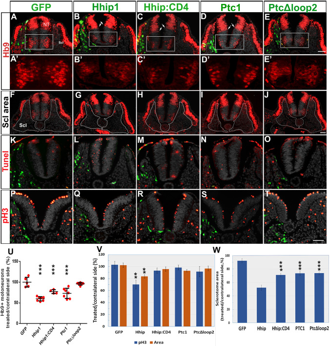
Fig. 2.
Electroporation of Hhip:CD4 and Ptc1 into the sclerotome inhibit motoneuron development without significantly affecting progenitor proliferation or survival. (A-E′) Electroporation (green) to the sclerotome followed by immunostaining for Pax7 and Hb9 1 day later. A′-E′ represent higher magnifications of the boxed regions in A-E. All plasmids, except PTCΔloop2, reduced the number of motoneurons adjacent to the transfected sclerotomes. Arrows in B-D show a slight ventralization of the ventral boundary of Pax7. (F-J) The same sections as in A-E in which the sclerotomes are delineated by white lines. (K-O) TUNEL staining (red). Weak immunostaining of electroporated plasmids (green) can be observed because no anti-GFP antibodies were used to enable better visualization of apoptotic nuclei. Only Hhip1 resulted in numerous TUNEL+ nuclei adjacent to the transfected sclerotome. Ectodermal staining reflects non-specific reactivity. (P-T) staining of mitotic nuclei with anti-pH3 (red). (U) Quantification of motoneurons in the hemi-NT facing the transfected mesoderm compared with control GFP. (V) Quantification of mitotic nuclei and area in the hemi-NT adjacent to the electroporated sclerotome. (W) Quantification of the relative area of electroporated sclerotomes based on sections such as those shown in F-J. NT, neural tube; Scl, sclerotome. Data are mean±s.e.m. **P<0.03, ***P<0.01. Scale bars: 50 µm.

