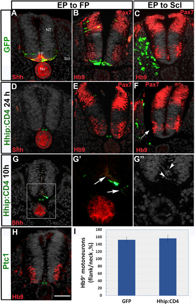
Fig. 8.

Sclerotome-derived Shh, but not FP-derived Shh, is necessary for motoneuron development. (A,B) Dorsoventral electroporation of control GFP showing (A) the presence of the labeled FP that co-expresses Shh protein. (B) Hb9+ motoneurons are dorsal to the labeled FP. (C) Hb9+ motoneurons in control GFP electroporation to the sclerotome. (D,E) Loss of FP tissue 24 h post-Hhip:CD4 electroporation. Asterisks in D,E denote the absence of a FP; D shows concomitant absence of Shh expression. Hhip:CD4 electroporation has no effect on ventral Hb9+ motoneurons or dorsal Pax7 expression (both in red). (F) In contrast, fewer motoneurons are apparent adjacent to the sclerotome transfected with Hhip:CD4 (arrow). (G-G″) Disintegration of the FP 10 h following Hhip:CD4 transfection. Note in G and G′ the almost complete absence of GFP signal in the FP domain (with only one GFP+ cell remaining), as well as the punctate expression of GFP (arrows) corresponding to disorganized and pyknotic nuclei (arrowheads in G″). (H) Loss of the FP upon electroporation of Ptc1 (green asterisks indicate the lack of an FP) shows no apparent effect on motoneurons. (I) Quantification of the proportion of Hb9+ motoneurons in flank (electroporated) and neck (intact region). Data are mean±s.e.m. No, notochord; NT, neural tube. Scale bar: 50 µm.
