Fig. 3.
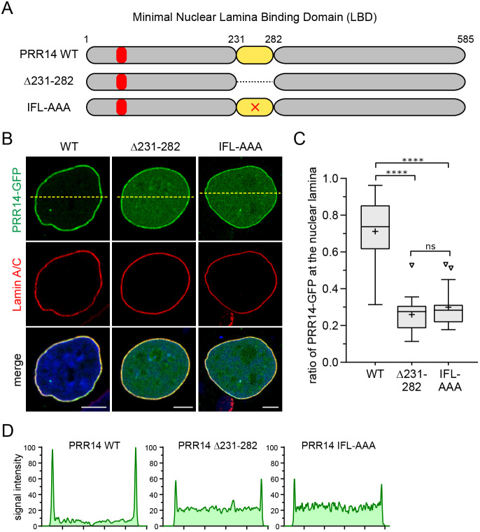
The 231–282 LBD is required for efficient localization of PRR14 to the nuclear lamina. (A) Diagram of human wild-type (WT) and mutated PRR14 depicting the 231–282 LBD, Δ231–282, and IFL to AAA substitution (×) in the conserved core sequence. The LAVVL HP1/heterochromatin-binding motif (52–56) is indicated in red. (B) Representative confocal images of HeLa cells transfected with the GFP-tagged PRR14 constructs (green) depicted in A, with anti-Lamin A/C staining (red) and DAPI counterstaining. Dashed lines indicate line sections for signal intensity profiles shown in D. (C) Box plot demonstrating the proportion of indicated PRR14–GFP protein at the nuclear lamina compared to the total nuclear signal, calculated using Lamin A/C signal as a mask. Boxes indicate the interquartile range with the median represented by a horizontal bar. Whiskers are drawn using the Tukey method and + indicates the mean value. n=20 cells per condition. (D) Line signal intensity profiles of sections indicated by dashed lines in B. ****P<0.0001; ns, not significant (one-way ANOVA with Dunn's multiple comparison test). Scale bars: 5 µm.

