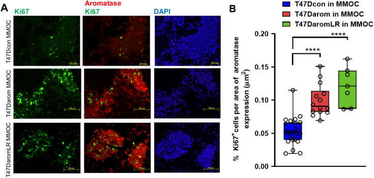
Fig. 5.
Immunofluorescence analysis of aromatase and Ki67 in BCa-MMOC section. (A) T47Dcon (top panel), T47Darom (middle panel) and T47DaromLR (bottom panel) were grown in BCa-MMOC and mammary gland sections and representative images were dually immunostained for Ki67 (in green) and aromatase (in red). All images were taken at 10× objective. (B) Quantitative analysis of the area of aromatase expression occupied by T47Dcon, T47Darom and T47DaromLR expressing aromatase (in red) within the mouse mammary gland. Results were quantitated and expressed as the percentage of proliferation (as measured by Ki67 positive cells µm−2 area occupied by the T47D variant cells expressing aromatase, ****P<0.0001). Five glands for each cell type were measured for aromatase and Ki67 staining.

