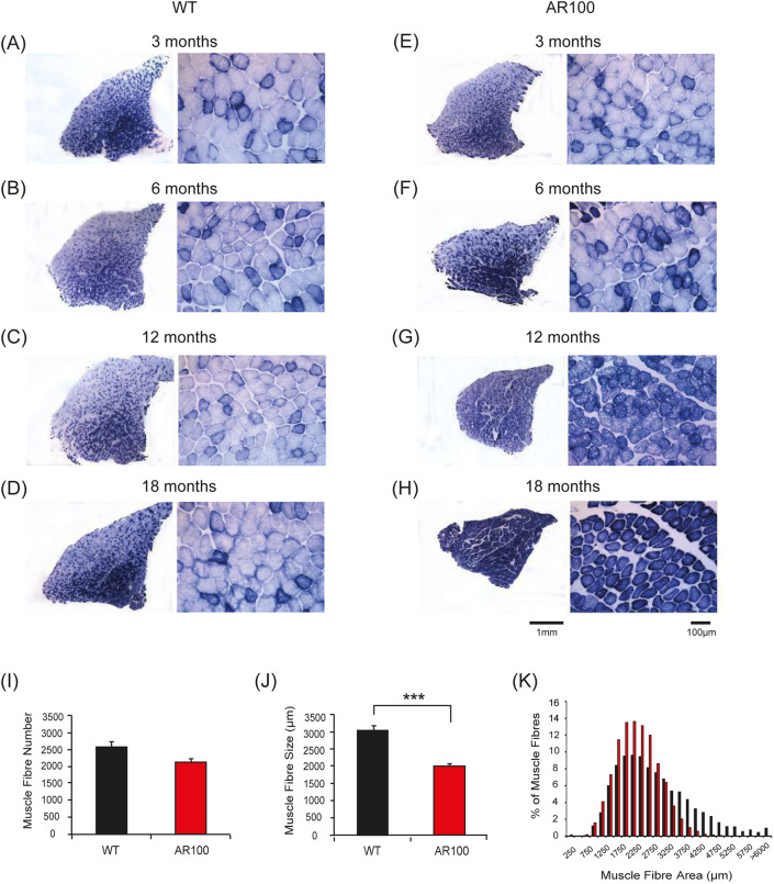
Fig. 4.
Increase in oxidative capacity of TA muscle in SBMA mice with disease progression. (A-H) Transverse TA muscle sections of WT (A-D) and AR100 (E-H) mice were stained for the mitochondrial respiratory enzyme SDH. (A-D) Muscle sections from WT mice at 3, 6, 12 and 18 months displayed a typical staining pattern, with intensely stained, highly oxidative fibres in the middle, and a characteristic mosaic pattern, representative of less oxidative, fast-twitch fibres in the outer region. (E) TA muscle of AR100 mice at 3 months of age was not different from that of WT mice. (F) At 6 months however, the first signs of muscle pathology were evident in AR100 mice. There appeared to be a slight increase in the number of intensely stained, oxidative fibres and evidence of muscle fibre grouping. (G) At 12 months of age, muscle fibre atrophy was clear and the number of intensely stained fibres was dramatically increased. (H) By 18 months of age the majority of fast-twitch fibres in AR100 TA muscle had been transformed to that of a slow-twitch, highly oxidative phenotype. Fibres were atrophied in comparison to those in WT TA muscles and muscle fibres were less densely packed, with increased space between them. (I) The number of muscle fibres present in AR100 mice was reduced but not significantly different to WT at 18 months of age. (J) At this late disease stage, the mean fibre area in TA of AR100 was significantly less than age-matched WT mice. (K) There was a clear reduction in larger size fibres accompanied by an increase in the number of smaller fibres. Statistical analysis was performed using Student's t-test (two-tailed) (n≥5 animals, ***P<0.001). Error bars represent s.e.m.

