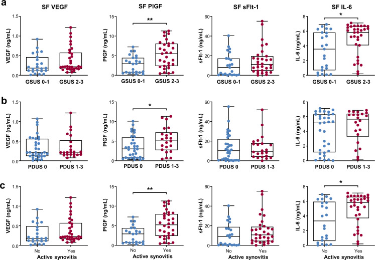
Fig. 2. Levels of VEGF, PlGF, sFlt-1, and IL-6 in the SF according to synovitis severity on US.
a VEGF, PlGF, sFlt-1, and IL-6 concentrations in the SF of RA patients with significant synovial hypertrophy (gray-scale US, GSUS 2 and 3) versus those with no or mild synovial hypertrophy (GSUS 0 and 1). b VEGF, PlGF, sFlt-1, and IL-6 concentrations in RA patients with increased vascularity (power-Doppler US, PDUS 1 to 3) and in those without (PDUS 0). c VEGF, PlGF, sFlt-1, and IL-6 concentrations in RA patients with active synovitis and in those without. Active synovitis was defined as GSUS ≥ 2 or PDUS ≥ 1. GSUS: gray-scale US; PDUS: power-Doppler US. Bars indicate the mean and SD. *p < 0.05, **p < 0.01 by Student’s unpaired t test.

