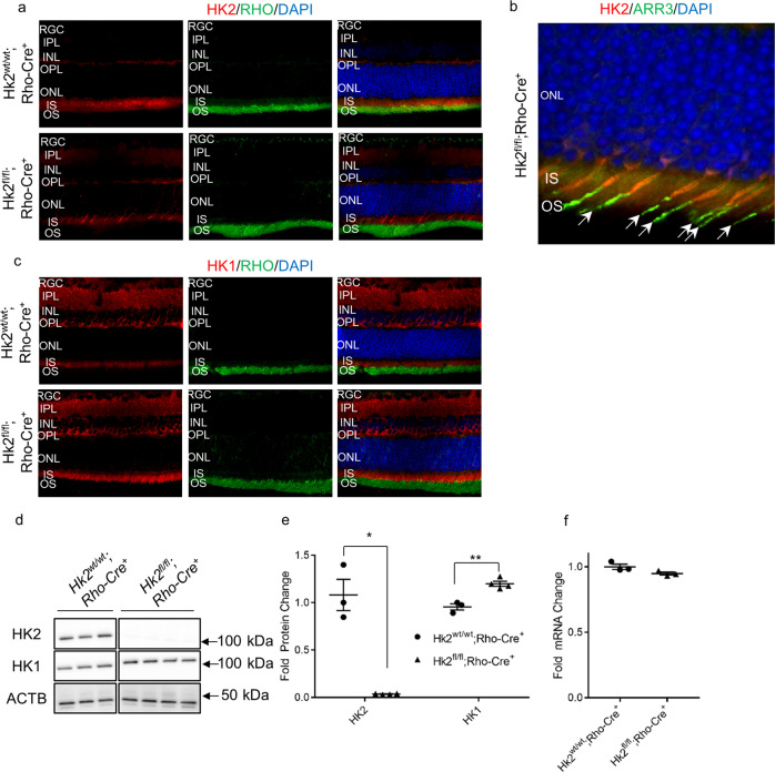
Fig. 2. Successful knockdown of HK2 in rod photoreceptors with compensatory upregulation of HK1.
a HK2 immunofluorescence (red) is found mainly in the inner segments of photoreceptors with little expression elsewhere in the retina of WT mice (Hk2wt/wt;Rho-Cre+). Rod photoreceptor-specific, Hk2 cKO mice (Hk2fl/lt;Rho-Cre+) lack expression of HK2 in the majority of photoreceptors. b Co-labeling of HK2 (red) and ARR3 (green) confirms that the remaining HK2 expression is limited to cone photoreceptors in cKO mice. c HK1 immunofluorescence (red) depicts expression mainly in the inner retina of WT mice while cKO mice show upregulation of HK1 in photoreceptor inner segments. Nuclei of retinal cells are stained with DAPI (blue). d Western blot demonstrating almost complete loss of HK2 in cKO mice with a compensatory upregulation of HK1. ACTB (β-actin) was used as a loading control. e Quantitative analysis of HK2 and HK1 protein levels shows a statistically significant decrease in the level of HK2 in cKO mice. HK1 protein levels are statistically significantly increased in the retinas of these animals as compared to WT animals. f Total Hk1 transcript levels are unchanged in cKO animals compared to WT animals. N = 3–4, *p < 0.05 **p < 0.01. RGC Retinal Ganglion Cell layer, IPL Inner Plexiform Layer, INL Inner Nuclear Layer, OPL Outer Plexiform Layer, ONL Outer Nuclear Layer, IS Inner Segments, OS Outer Segments, DAPI 4′,6-diamidino-2-phenylindole, RHO Rhodopsin, ARR3 Cone Arrestin.

