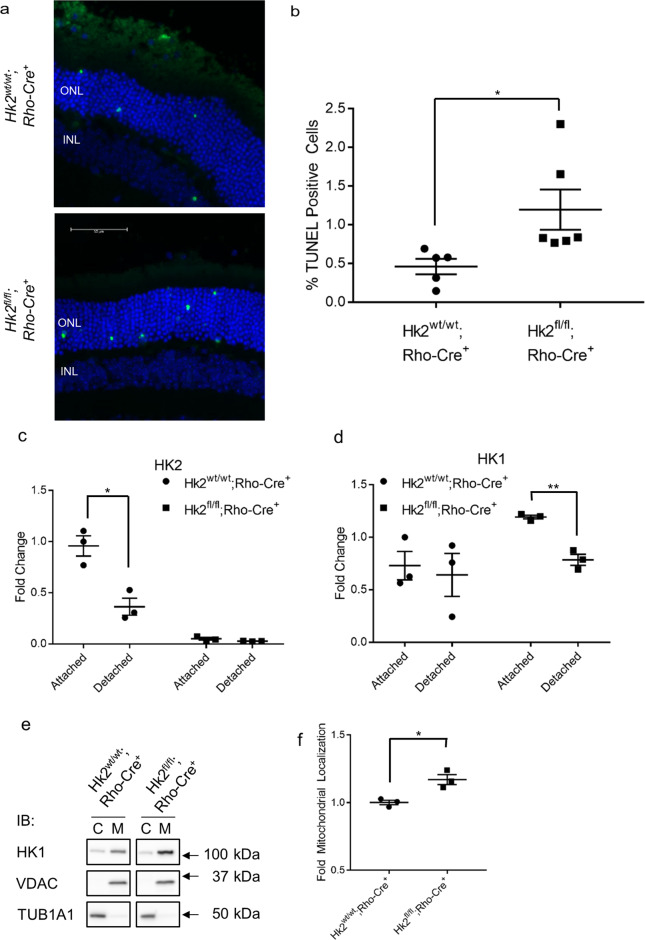
Fig. 3. HK2 regulates photoreceptor survival after retinal detachment.
a Representative images of TUNEL-stained photoreceptors (green) in detached regions of mouse retina 3 days post-retinal detachment. Nuclei of cells within ONL (outer nuclear layer) and INL (inner nuclear layer) are stained with DAPI (blue). b Quantification of TUNEL-positive cells in the ONL. c Quantitative analysis of HK2 protein levels 3 days after retinal detachment. HK2 is down-regulated following retinal detachment in WT mice. d Quantitative analysis of HK1 protein levels 3 days after retinal detachment. HK1 is downregulated following retinal detachment in cKO mice only. e Representative Western blot for data quantified in f. f Total HK1 protein levels are significantly enriched in the mitochondrial fraction in cKO 3-days post-retinal detachment in cKO animals versus WT animals. N = 3–6 animals per group, *p < 0.05, **p < 0.001.

