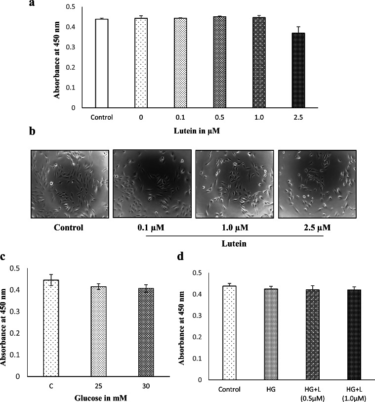
Fig. 2.
Effect of lutein and glucose on the viability and morphology of ARPE-19 cells. a The cell viability of ARPE-19 treated with lutein at the noted concentrations (0, 0.1, 0.5, 1.0 & 2.5 μM) for 24 h was analyzed by WST-1 assay. Values are mean ± SD (n = 3). b The cell morphology of ARPE-19 treated with lutein for 24 h was observed under a phase-contrast microscope, and the representative picture is presented. c The cell viability of ARPE-19 treated with glucose at the different concentrations (25 & 30 mM) for 24 h was analyzed by WST-1 assay. Values are mean ± SD (n = 3). d The effect of lutein (L-1 μM) on the viability of ARPE-19 cells treated with high-glucose (HG-25 mM) was analyzed by WST-1 assay. Values are mean ± SD (n = 3)

