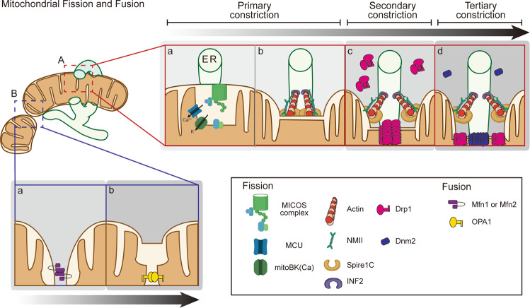
Fig. 1. Mechanism of mitochondrial fission and fusion.
a A mitochondrion is divided via multiple constrictions of the OMM and IMM. A The majority of mitochondrial fission takes place at contact sites between the mitochondria and the ER, which act as platforms for the accumulation of various proteins, such as actin, ion channels, and motor proteins, to initiate the constriction of the OMM and/or IMM. INF2 and spire1C induce actin polymerization on the ER side and mitochondrion side, respectively, allowing for the generation of an MNII-mediated pulling force. B IMM constriction proceeds independent of actin polymerization and Drp1 activation. It is required for the MCU-dependent Ca2+ influx that destabilizes the mitochondrial inner structure. Ca2+ influx activates mitoBK(Ca), which induces mitochondrial bulging. C Cytosolic Drp1 forms a ring structure at the constricted site, which further constricts the mitochondrion by GTP hydrolysis. D Dnm2 finally constricts the mitochondrion to a diameter of <10 nm, which is sufficient for lipid autocleavage. b Mitochondrial fusion is mediated by dynamin-related proteins, such as Mfn1/2 and OPA1. When two mitochondria approach each other, Mfn docking is initiated. The interaction between Mfns in their non-GTP binding state reduces the gap between the mitochondria. Then, GTP hydrolysis drives membrane fusion. Because OPA1 interacts with Mfns, it may promote IMM fusion following OMM fusion. Oligomerization and GTP hydrolysis promote conformational changes that bring the two membranes into close proximity, facilitating IMM fusion.

