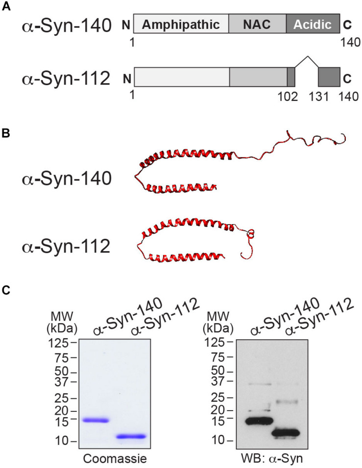
FIGURE 1.
α-Syn-112 splice isoform. (A) Domain diagram of α-syn-140 and α-syn-112 showing the result of exon 5 deletion. (B) (top) The NMR structure of human α-syn-140 bound to lipid micelles (Ulmer et al., 2005), and (bottom) predicted structure of α-syn-112 (UCSF Chimera software). α-Syn-112 is predicted to have a slightly extended alpha helix. (C) Coomassie-stained SDS-PAGE gel and Western blot showing the size difference between α-syn-140 (14 kDa) and α-syn-112 (12 kDa). In the Western blot, the higher molecular weight bands represent minor fractions of dimeric α-syn-140 and α-syn-112.

