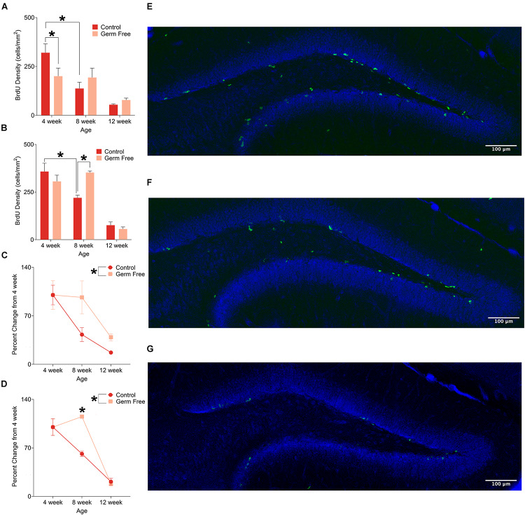
FIGURE 2.
(A) Mean (± SEM) BrdU-positive cells in the DG of male germ-free and control mice. Control mice show a clear pattern of age-related decline in cell proliferation, whereas germ-free mice show no such reduction between 4 and 8 weeks of age. Germ-free mice also show reduced cell proliferation relative to controls at 4 weeks of age. (B) Mean (± SEM) BrdU-positive cells in the DG of female germ-free and control mice. As with males, female control mice show an age-related decline in cell proliferation with this effect being absent in germ-free mice between 4 and 8 weeks of age. Moreover, female germ-free mice have increased cell proliferation relative to controls at 8 weeks of age. (C) Mean (± SEM) cell proliferation in males depicted as percent-change from the baseline (4 week old) number of BrdU-positive DG cells. Cell proliferation remains abnormally elevated in germ-free mice as they age relative to controls (D). Mean (± SEM) cell proliferation in females depicted as percent-change from the baseline (4 week old) number of BrdU-positive DG cells. As with male germ-free mice, cell proliferation in female germ-free mice remains abnormally elevated relative to controls particularly at 8 weeks of age. (E–G) Representative photomicrographs of BrdU-positive cells (green) and propidium iodide (blue) in the DG of 4 week old (E), 8 week old (F), and 12 week old (G) control mice illustrating the age-related decrease in cell proliferation. Control male 4 week n = 10, 8 week n = 7, 12 week n = 8. Control female 4 week n = 4, 8 week, n = 5, 12 week n = 4. Germ-free male 4 week n = 5, 8 week n = 7, 12 week n = 8. Germ-free female 4 week n = 4, 8 week, n = 3, 12 week n = 3. *p < 0.05.

