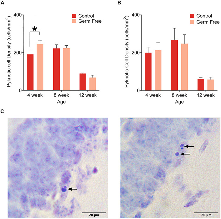
FIGURE 3.
(A) Mean (± SEM) pyknotic cells in the DG of male germ-free and control mice. The number of pyknotic cells is highest in 4 week old animals and lowest in 12 week old animals. Additionally, germ-free mice show an increased rate of cell death at 4 weeks of age. (B) Mean (± SEM) pyknotic cells in the DG of female germ-free and control mice. As is the case in male mice, the rate of cell death is highest in 4 week old animals and lowest in 12 week old animals. There is also a slight trend toward elevated cell death at 8 weeks of age. In contrast to male mice, female germ-free mice showed no change in the rate of cell death relative to controls. Control male 4 week n = 10, 8 week n = 10, 12 week n = 9. Control female 4 week n = 4, 8 week, n = 4, 12 week n = 4. Germ-free male 4 week n = 9, 8 week n = 9, 12 week n = 10. Germ-free female 4 week n = 4, 8 week, n = 4, 12 week n = 4. (C) Representative photomicrographs of DG cells with pyknotic morphology. *p < 0.05.

