Figure 5.
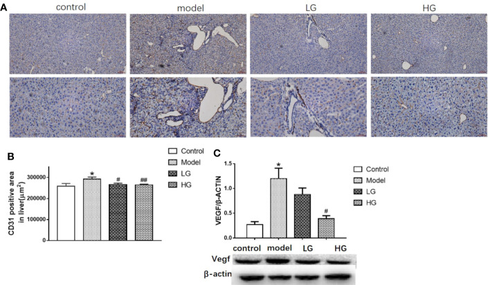
Angiogenic ability of hepatocellular carcinoma in microenvironment is inhibited by GHJCD. (A) Representative image of CD31 immunohistochemical staining in liver tissue. Top scale bar: 200 µm; bottom scale bar: 100 µm. (B) Area of CD31-positive distribution in liver tissue (µm2). (C) Expression of VEGF in liver tissue. CD31, VEGF data in the liver cancer microenvironment were analyzed by ANOVA. The homogeneity test was first performed, P > 0.05, the variances were uniform, and the LSD (L) test was used for multiple comparisons afterward, P < 0.05, the variances were not uniform.*p < 0.05, **p < 0.01 vs. control; #p < 0.05 vs. model.

