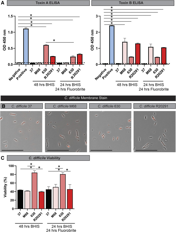
Fig. 2.
Clostridioides difficile strains secrete toxins, maintain shape and viability after incubation in Fluorobrite DMEM. A: toxin A and B ELISAs with C. difficile strains 37, M28, 630, and R20291 after 48 h in BHIS or 24-h incubation in BHIS and an additional 24 h incubation in Fluorobrite DMEM. ELISAs were examined on a plate reader at optical density (OD) of 450 nm. B: FM 4–64 membrane staining in C. difficile 37, M28, 630, and R20291 after 24-h incubation with BHIS and an additional 24 h incubation in Fluorobrite DMEM. Scale bar = 50 μm. C: cell viability as determined by BacLight live/dead cell staining. Cell staining was examined at excitation: 485 nm/emission: 530 nm (live) and excitation: 485 nm/emission: 630 nm (dead) on a Synergy H1 Microplate Reader, and viabilities were calculated by dividing the fluorescence intensity of 485/530 by the fluorescence intensity at 485/630 × 100%.

