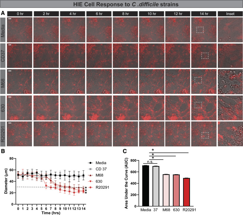
Fig. 3.
Human intestinal enteroids (HIEs) exhibited delayed cell rounding in response to toxigenic Clostridioides difficile strains. HIEs derived from the jejunum were transduced with the LifeAct-Ruby sensor, which labels F-actin with red fluorescent protein. Cell rounding was visualized by live cell microscopy on a Nikon TiE with a ×20 Plan Apo (NA 0.75) differential interference contrast objective, using a SPECTRA X LED light source and ORCA-Flash 4.0 sCMOS camera (scale bar, 50 µm). A: representative images of LifeAct-Ruby HIEs over time (1–16 h) after exposure to Fluorobrite DMEM medium alone or conditioned Fluorobrite from C. difficile strains CD37, M68, 630, or R20291. Insets indicate significant rounding occurs with toxigenic C. difficile strains (M68, 630, and R20291), but this cell rounding occurs at later time points. B: FIJI (formerly ImageJ) software was used to define cell membranes (as denoted by actin labeling) and cell diameter over time. C: resulting curves were assessed for the area under the curve (AUC), which demonstrates decreased cell diameter with M68, 630, and R20291. *P < 0.05, 1-way ANOVA; n = 3 independent experiments, n = 6/experiment. NS, not significant.

