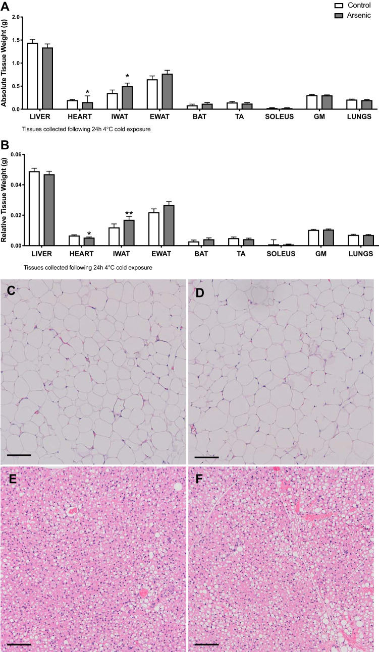
Fig. 4.
Chronic arsenic exposure increased inguinal white adipose tissue (iWAT) relative and absolute tissue weight. Absolute (A) and relative tissue weight (B) (n = 13 arsenic vs. n = 13 controls). Representative hematoxylin and eosin (H&E) staining (×10) depicting control iWAT (C); arsenic-treated iWAT (D); control brown adipose tissue (BAT) (E); arsenic-treated BAT (F); n = 10; 5 arsenic vs. 5 controls. Data are represented as least squares means ± SE, with statistical significance determined by linear mixed models accounting for cohort as a random effect. **P < 0.01; *P < 0.05 arsenic vs. controls. Scale bar = 100 μM. eWAT, epididymal WAT; TA, tibilias anterior; GM, gastrocnemius muscle. Tissues were collected following a 24-h 4°C cold exposure. No statistical differences in pancreas absolute or relative weight were observed (data not shown).

