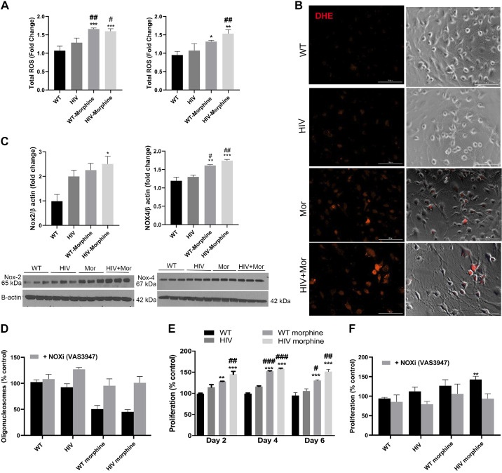
Fig. 7.
Increased oxidative stress in endothelial cells isolated from HIV-transgenic (HIV-Tg) rats administered with morphine. A: RPMECs (3,000 cells/well) were plated on a 96-well plate, and, at 80% confluency, cells were serum starved using 0.5% serum-containing media for 3 (left) and 12 (right) h followed by DCF assay. ROS, reactive oxygen species; WT, wild type. B: for DHE staining, confluent rat endothelial cells were serum starved for 3 h followed by incubation with DHE dye (5 nM) for 30 min. The cells were then fixed with 4% paraformaldehyde, and images were captured using a Lion Heart imaging station (scale: 100 µm). Mor, morphine. C: confluent RPMECs were serum starved for 3 h followed by Western blotting for NADPH oxidase 2 (NOX2) and NOX4 expression. All the respective blots were stripped and reprobed for β-actin antibody used as loading control. NOX2 and -4 were run with the same samples on different gels due to similar molecular weight. D: RPMECs were serum starved 24 h after plating of cells in a 96-well plate followed by cell death ELISA at day 2 posttreatment in the presence or absence of VAS3947. E and F: MTS proliferation assay at days 2, 4, and 6 (E) and at day 4 (F) posttreatment in the presence or absence of VAS3947. Values are means ± SE of n = 3 rats/group. **P < 0.01, *P < 0.05, ***P < 0.001 vs. WT. ##P < 0.01, #P < 0.05, ###P < 0.001 vs. HIV.

