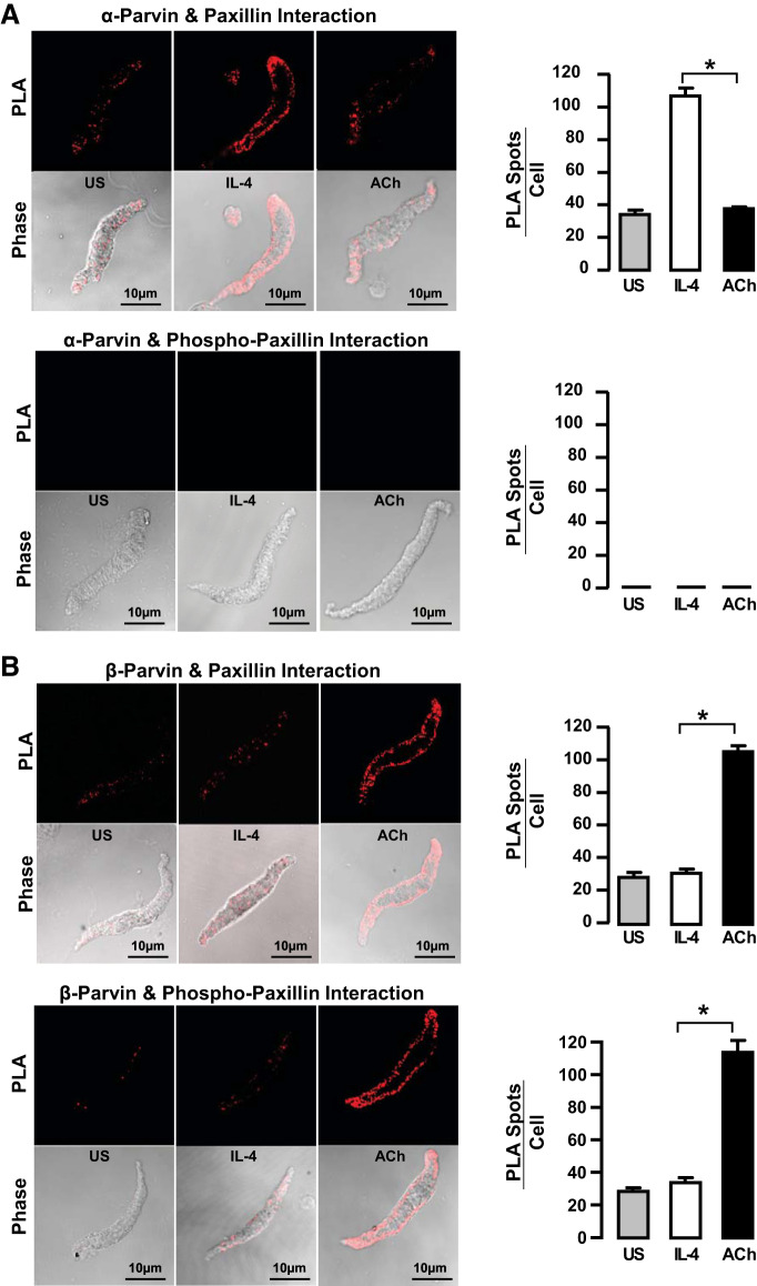
Fig. 9.
IL-4 promotes interactions between α-parvin and unphosphorylated paxillin, whereas ACh promotes interactions between β-parvin and phosphorylated paxillin in freshly dissociated tracheal smooth muscle cells. Proximity ligation assay (PLA) fluorescence images show the interaction of α-parvin or β-parvin with paxillin or pY118-paxillin in dissociated cells stimulated with ACh or IL-4 or unstimulated (US). A: significantly more paxillin interacts with α-parvin in cells stimulated with IL-4 than in cells stimulated with ACh or unstimulated cells. Paxillin that interacts with α-parvin is not phosphorylated. B: significantly more β-parvin interacts with paxillin in cells stimulated with ACh than in cells stimulated with IL-4 or unstimulated cells. Paxillin that interacts with β-parvin is phosphorylated. All values are means ± SE. *Significant difference between IL-4- and ACh-stimulated groups (n = 10–20 cells per group). Statistical analysis by one-way ANOVA. P < 0.05 was considered significant.

