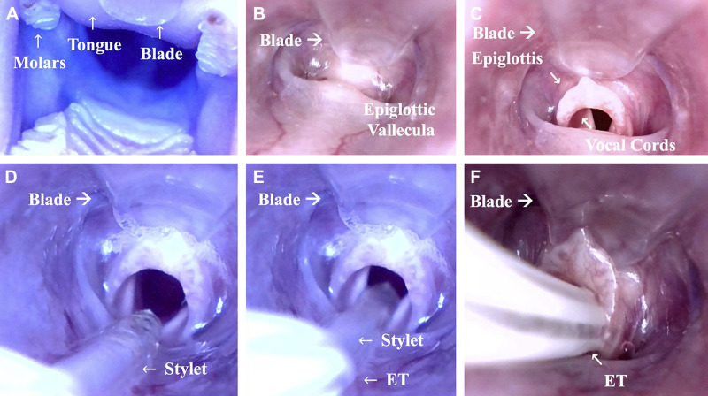
Fig. 2.
Insertion of the video laryngoscope (VL) and field of view during intubation. The VL is introduced into the mouth of the rat with the blade gliding along the tongue (A). The tip of the blade is carefully placed next to the epiglottic valleculae (B). The epiglottis is raised with a slight move of the tip of the blade in ventral direction (C). The stylet is inserted into the glottis (D). Once the stylet has passed the glottis ~1 cm (E), the endotracheal tube (ET) is passed over the stylet into the trachea (F), and finally the stylet is pulled back. The procedure can also be watched in Supplemental Video S1 available at www.doi.org/10.6084/m9.figshare.11891571.v2.

