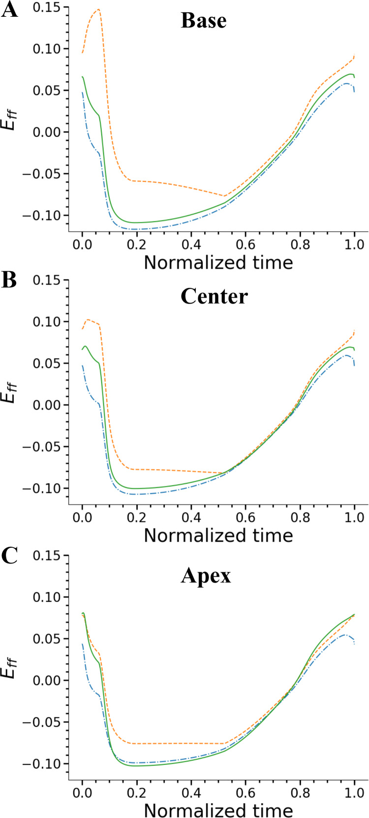
Fig. 4.
Fiber strains in the left ventricle (LV) as a function of the cardiac cycle time at 3 transmural locations of base (A), center (B), and apex (C) of the LV. At each axial location, 3 transmural regions of subendocardium (solid line), midwall (dashed line), and subepicardium (dash-dotted line) regions were sampled. In all the axial regions, transmural difference is significantly higher at peak systole. Eff, myofiber strain.

