Fig. 2.
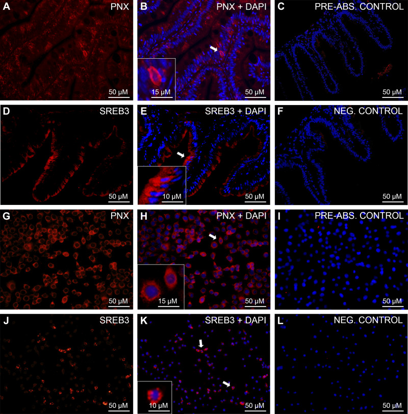
Immunohistochemical analysis of phoenixin (PNX)-like and super conserved receptor expressed in brain 3 (SREB3)-like in zebrafish gut and zebrafish liver (ZFL) cells. Shown are representative sections of zebrafish gut (A–F) and ZFL (G–L) cells showing PNX-like (red; A–B and G–H) and SREB3-like (red; D–E and J–K) immunoreactivity (ir). No PNX-/SREB-3-like ir was observed in either preabsorption (C and I) or secondary antibody-alone negative control (F and L). Nuclei are shown in blue (DAPI). Scale bars are indicated in each image.

