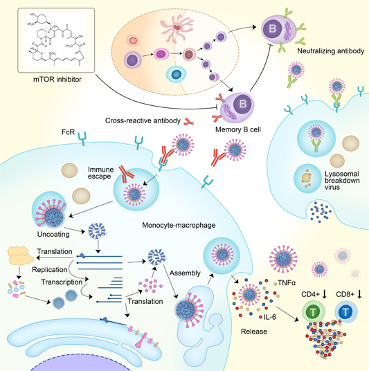
Figure 1.

Antibody‐dependent enhancement (ADE) and cytokine release during coronavirus infection The upper right part of the figure: B cells differentiate and mature from their progenitor cells in the germinal center, and finally become plasma cells, secreting IgM at an early stage and IgG about 2 weeks post‐infection. These IgGs are neutralizing antibodies that bind to coronavirus surface spike proteins. The constant region of the antibody binds to the Fc receptor on the surface of the monocyte‐macrophage and is engulfed into the cell. Then, the lysosome recognizes the antibody‐virus complex, and the phagocytic complex is decomposed. Finally, virus fragments are released as part of the normal immune process. The lower left part of the figure: The antibodies secreted by memory B cells are cross‐reactive antibodies, which will inhibit the secretion of neutralizing antibodies by plasma cells. The cross‐reactive antibody binds to the virus with weak affinity, and after the complex binds to the Fc receptor, it is engulfed by monocyte macrophages. After entering the monocyte‐macrophage, because of weak affinity, the virus is separated from the cross‐reactive antibody and is not engulfed by the lysosome, leading to the immune escape. Subsequently, the virus uncoates, replicates, and assembles. Finally, a large number of mature viruses are released from the monocyte macrophages. At the same time, IL‐6, TNF‐α, and other cytokines are also released. These cytokines downregulate T cells (including CD4+ and CD8+). With more and more cytokines released, a so‐called cytokine storm is formed during the ADE process. The upper left corner shows that an mTOR inhibitor, rapamycin, could inhibit the activation of memory B cells and therefore inhibit the ADE process. IgG, immunoglobulin G; IgM, immunoglobulin M; IL‐6, interleukin 6; mTOR, mammalian target of rapamycin; TNF‐α, tumor necrosis factor α
