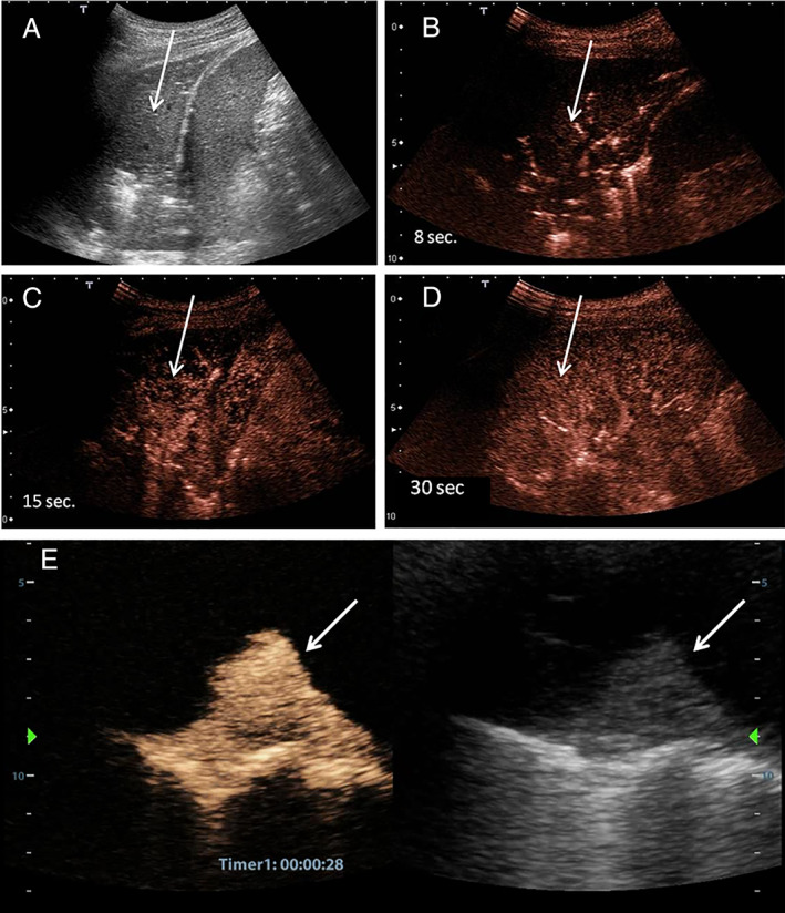
Figure 1.

A, Ultrasound image of left basal pneumonia (arrows). The parenchyma of the lower lung lobe is consolidated. B, Contrast‐enhanced US 8 seconds from the injection of 2.4 mL of SonoVue. Arterial enhancement with a segmental appearance is shown. C, Initial parenchymal phase. D, Perfusional parenchymal phase. The whole lobe is enhanced. E, Right, Ultrasound image of compressive pulmonary atelectasis. Left, Same image at 30 seconds from the injection of 2.4 mL of SonoVue. The compressed and airless parenchyma is strongly enhanced.
