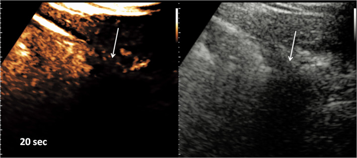
Figure 3.

Detail of a small cuneiform subpleural consolidation (arrows). Contrast‐enhanced US (left) and grayscale B‐mode (right) images were acquired at 20 seconds from the injection of 2.4 mL of SonoVue. An obvious defect of perfusion of the lesion is shown.
