
Figure 2.
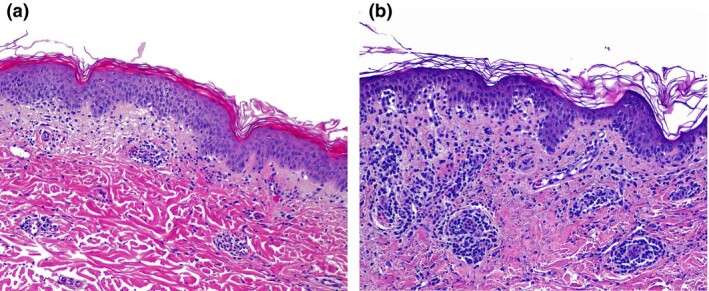
(a) Histological examination of Patient 1 showed normal basketweave stratum corneum, mild to moderate spongiosis and lymphocytic exocytosis in the epidermis, while the dermis showed dilated vessels filled with neutrophils, extravasation of red blood cells, and lymphocytic perivascular and interstitial infiltrate. (b) Patient 3 had basal vacuolar changes with interface dermatitis.