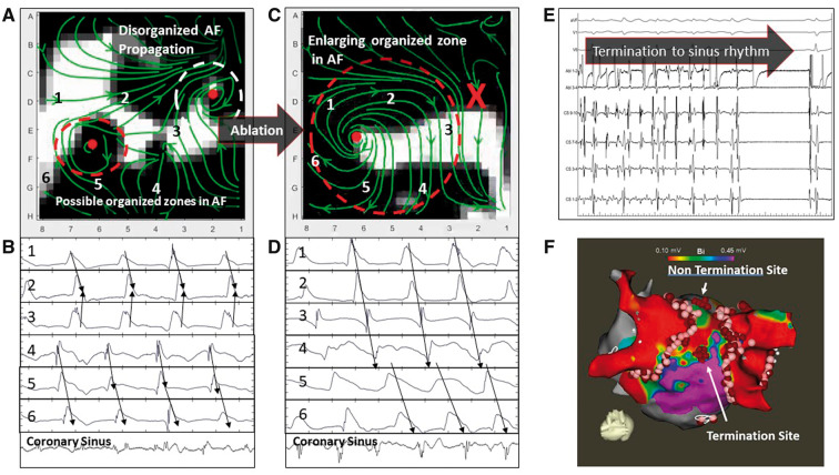
Figure 2.
WFF of persistent AF in a 55-year-old man with termination to sinus rhythm. (A) WFF and corresponding electrograms. WFF shows two organized areas (coloured ellipses) separated by disorganization. Unipolar electrograms at precise points marked on WFF streamlines confirm 1:1 activation near the red ellipse, within disorganized activity seen on bipolar CS electrograms. The white ellipse area was ablated, but AF did not terminate. (B) WFF after ablation of white organized ellipse (marked X), shows enlargement of the residual organized area (red ellipse). Unipolar electrograms confirm 1:1 activation within the red ellipse. (C) Ablation at the centre of this primary area terminated AF to sinus rhythm. (D) Electroanatomic map. AF, atrial fibrillation; CS, coronary sinus; WFF, wavefront field.

