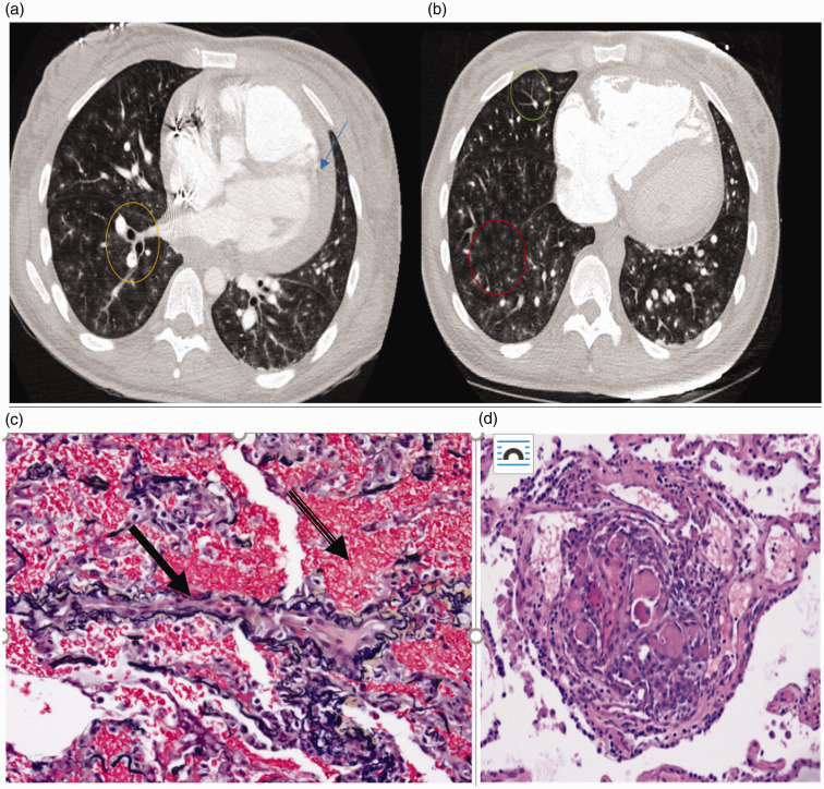
Fig. 1.
(a and b) Case 2: Axial images from computed tomography angiogram demonstrating diffuse bilateral centrilobular ground glass opacities (red circle), tree in bud opacities (green circle), central peribronchovascular interstitial thickening (yellow circle), and pericardial effusion (blue arrow). Case 2. (c) Elastic fiber (VVG)-stained section of lung showing a vein (solid arrow) with marked fibrous intimal thickening and almost complete obliteration of the lumen and extensive alveolar hemorrhage (banded arrow). (d): H&E-stained section of the lung showing a plexiform lesion present adjacent to two pulmonary artery branches composed of several slit-like lumens with prominent cellularity and fibrin thrombi.

