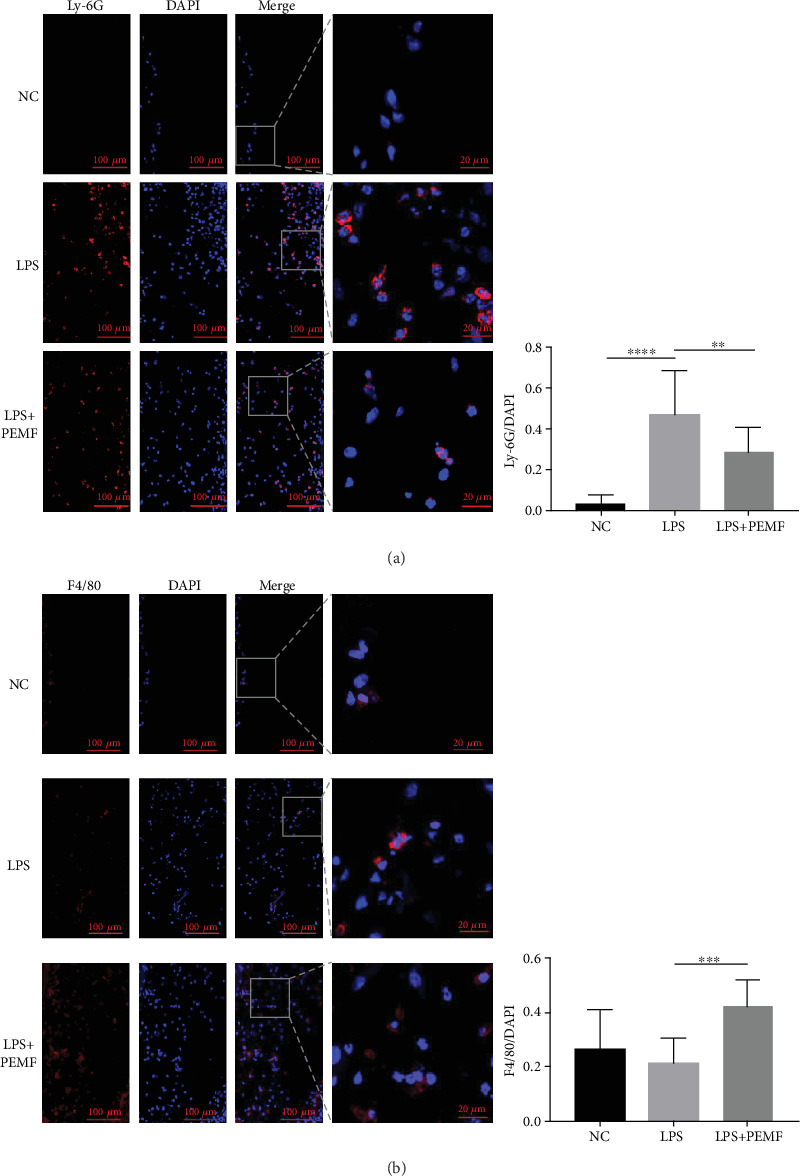
Figure 3.

The proportion of inflammatory cells was changed after PEMF treatment. After LPS was used to induce inflammation in the air pouch, the experimental group was given PEMF treatment; a small piece of skin from the air pouch was sampled for immunofluorescent staining. (a) Immunofluorescence showed a decrease in Ly-6G-labelled neutrophils after PEMF treatment. Statistical analysis was performed by one-way ANOVA, and multiple comparisons were performed using Tukey's test. Error bars represent the mean ± SD of fifteen independent experiments. ∗∗P < 0.01, ∗∗∗∗P < 0.0001. (b) Immunofluorescence showed an increase in macrophages after PEMF treatment. F4/80-labelled macrophages. Statistical analysis was performed by one-way ANOVA, and multiple comparisons were performed using Tukey's test. Error bars represent the mean ± SD of at least eight independent experiments. ∗∗∗P < 0.001.
