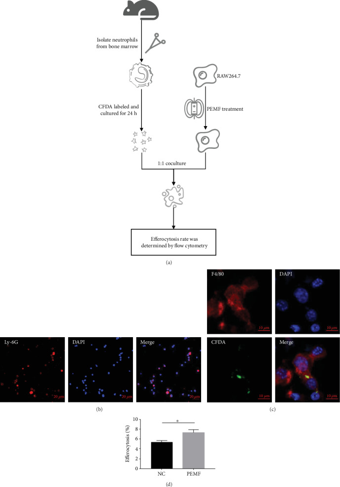
Figure 4.

The efferocytosis in macrophages is increased by PEMF treatment. (a) The procedure of efferocytosis: neutrophils were isolated from the bone marrow and cultured for 24 hours after CFDA labelling to induce apoptosis. The RAW264.7 cells treated with PEMF for 1 hour were 1 : 1 cocultured with apoptotic neutrophils for 4 hours. The efferocytosis rate was measured by flow cytometry. (b) Neutrophil purity was detected by immunofluorescence after isolation of mouse bone marrow neutrophils. (c) A confocal microscope was used to observe the efferocytosis. (d) The percentage of efferocytosis was measured by flow cytometry. Statistical analysis was performed by t test. Error bars represent the mean ± SD of three independent experiments. ∗P < 0.05.
